
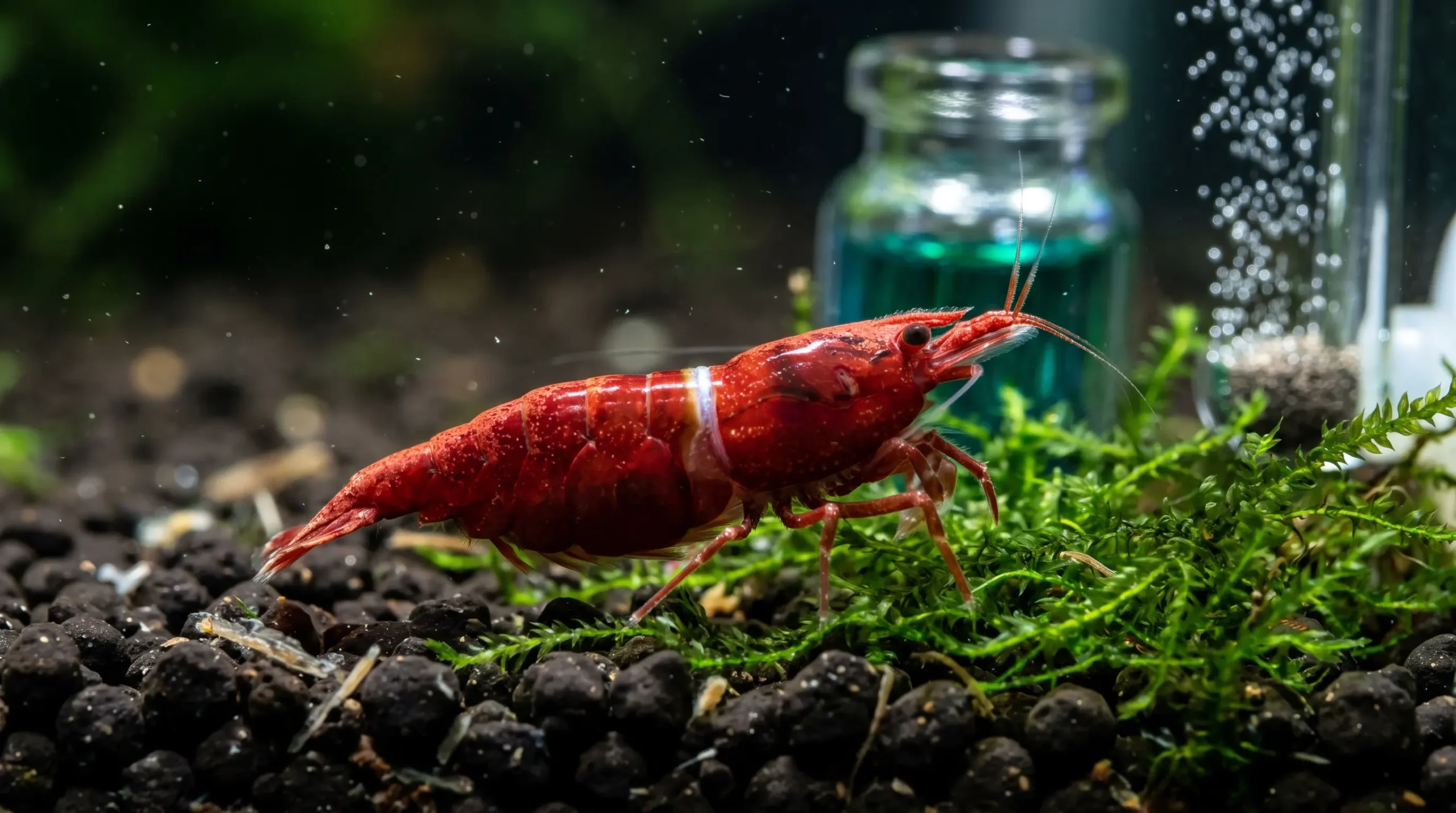
Few events in freshwater invertebrate keeping trigger as much immediate panic as discovering a white ring of death cherry shrimp casualty inside a thriving aquarium. When hobbyists spot a stark, pale translucent band splitting the carapace behind a shrimp’s head, community forums frequently declare an unavoidable death sentence. This pervasive fear drives cultivators to search frantically for cherry shrimp molting failure causes and fix protocols to prevent a catastrophic colony die-off. Understanding whether this visible separation represents normal cuticular apolysis or a fatal incomplete molt is essential for maintaining stable dwarf shrimp populations.
In standard freshwater setups, solving these molting failures requires looking beyond generic water hardness guidelines and examining the delicate decapod endocrine cascade. Decapod crustaceans like Neocaridina davidi must periodically undergo ecdysis, a complex physiological process tightly coordinated by Y-organ ecdysteroids and endogenous mineral salvage within stomach gastroliths. When ambient General Hardness (GH) deviates or osmotic shock occurs, this synchronized mechanical shedding fails. Identifying the distinct biochemical factors that govern cuticle shedding allows cultivators to rescue compromised stock and eliminate molting mortality permanently.
The primary catalyst for mid-molt fatalities typically stems from unmanaged water chemistry imbalances, including evaporative TDS creep, rapid hypo-mineralized water changes, and skewed calcium-to-magnesium (Ca:Mg) ratios. Furthermore, housing hard-water dwarf shrimp over active buffering aquasoils strips critical Carbonate Hardness (KH), exhausting cellular ATP reserves required for the physical molt jump. By implementing steady drip-refill methods, maintaining a verified 3:1 to 2:1 Ca:Mg ratio, and providing bioavailable dietary sterols, hobbyists can successfully resolve white ring of death cherry shrimp mortalities and ensure flawless ecdysis across every breeding cycle.
Table of Contents
Competitor Authority Analysis and Scientific Content Gaps: Unmasking the White Ring of Death Cherry Shrimp
Evaluation of Existing Ornamental Shrimp Literature and Molting Diagnostics
A systematic review of high-authority ornamental crustacean publications reveals polarized and scientifically incomplete coverage of the white ring of death cherry shrimp pathology. Mainstream blogs frequently identify surface correlations between water hardness and shrimp mortality but propagate conflicting diagnostic rules. Several prominent care guides assert that any visible white separation line behind a shrimp’s head carapace represents an irreversible death sentence.
Conversely, empirical tracking of live specimens demonstrates that normal pre-molt apolysis often presents an identical visual band prior to successful shedding. Existing commercial guides rarely connect home titration drop tests with peer-reviewed decapod physiology. Consequently, they overlook how intracellular calcium transport, amorphous mineral precursors, and branchial acid-base energetics govern cuticular shedding and postmolt calcification.
Understanding actionable cherry shrimp molting failure causes and fix strategies requires evaluating what leading hobbyist resources provide alongside the critical biochemical gaps they leave unresolved:
| Authority Domain / Source | Core Strengths Covered | Limitations & Unaddressed Technical Gaps |
|---|---|---|
| Aquarium Breeder | Explains the mechanical U-bend failure, debunks the excess dietary protein myth (citing 40% to 47% crude protein tolerances), and states ideal 3:1 Ca:Mg ratios. | Does not provide stoichiometric equations to calculate dissolved Mg2+ from GH/Ca titration tests, nor does it address the 24-hour pre-molt visual prediction window. |
| Shrimply Explained | Provides empirical 20-case survival tracking on pre-molt rings (showing 60% survival even with ≥ 2 mm breaks and scrunched edges) alongside Ca:Mg ratio math. | Lacks deep biochemical integration with Carbonate Hardness (KH), carbonic anhydrase upregulation, active soil cation stripping, and ACC glycolytic stabilization. |
| ShrimpKeepers & BucePlant | Clear beginner checklists on target General Hardness bounds (6 to 12 dGH), reverse osmosis remineralization, plant pesticide quarantine, and drip acclimation basics. | Falsely states that any shrimp showing a visible white ring is doomed, overlooking normal apolysis posture breaks and post-mortem vs. pre-molt clinical distinctions. |
| Gensou & TheShrimpFarm | Details neuroendocrine X-organ/Y-organ ecdysone cascades, endogenous gastrolith calcium reabsorption (20% to 50%), and continuous overflow water change systems. | Recommends potassium iodide dosing without warning that standard test kits cannot measure iodine and that overdosing induces acute decapod toxicity. |
| Shrimp Tank 101 & Peqaboo | Effectively debunks marine-derived liquid iodine myths in freshwater aquariums and isolates delayed 12-to-48-hour post-water-change mortality clusters. | Omits the molecular role of magnesium ions in modulating cuticular alkaline phosphatase activity and synthesizing structural magnesium-calcite in the endocuticle. |
Six Unaddressed Biochemical and Diagnostic Content Gaps
Bridging the divide between basic hobbyist observation and decapod endocrinology requires addressing six critical technical content gaps absent from competing literature. First, visual ring diagnostics must separate benign posture-dependent apolysis gaps from high-risk scrunched-edge fractures using empirical 24-hour resolution data. Tracked case data confirms that 60% of live specimens showing severe breaks still complete ecdysis safely.
Second, standard guides treat General Hardness (GH) as a monolithic number. Peer-reviewed crustacean biology demonstrates that magnesium (Mg2+) directly modulates alkaline phosphatase (ALP) activity and co-precipitates as structural magnesium-calcite; without sufficient magnesium, calcium deposition stalls even in mineral-dense water.
Third, hobbyist articles rarely explain how freshwater Neocaridina davidi salvage 20% to 50% of old cuticular calcium into paired stomach gastroliths 7 to 10 days before molting. They stabilize this reserve as Amorphous Calcium Carbonate (ACC) using matrix phosphoproteins like GAP65 and glycolytic intermediates like phosphoenolpyruvate (PEP) for immediate postmolt mobilization.
Fourth, nutritional guides overlook that crustaceans cannot synthesize sterol rings de novo. They require dietary cholesterol as the biochemical precursor for Y-organ ecdysteroid synthesis, while starving colonies of protein to prevent mythical overgrowth triggers cannibalism and strips the cuticle of its structural protein matrix.
Fifth, housing Neocaridina over active buffering substrates or allowing Carbonate Hardness (KH) to drop below 2 dKH forces branchial ionocytes to upregulate Carbonic Anhydrase (CA) enzymes. This drains cellular ATP required to power the physical molt jump.
Finally, aquarists must be cautioned against blind liquid iodine dosing and chronic sodium bicarbonate (NaHCO3) additions, which induce cumulative sodium toxicity compared to potassium bicarbonate (KHCO3) or aragonite buffering. Resolving these technical gaps equips breeders to manage white ring of death cherry shrimp risks systematically.
Anatomical and Mechanical Pathology of the White Ring of Death Cherry Shrimp
Encountering a white ring of death cherry shrimp casualty represents one of the most common yet frequently misdiagnosed emergencies in freshwater invertebrate keeping. Because dwarf shrimp (Neocaridina davidi) are encased in a rigid, non-expanding chitin-protein and calcium carbonate exoskeleton, somatic growth and ovarian maturation require periodic shedding and reconstruction of the entire outer cuticle—a process known scientifically as ecdysis.
In rapidly growing juvenile Neocaridina, ecdysis occurs every 7 to 14 days, whereas sexually mature adults maintained at 22°C to 26°C (72°F to 78°F) molt every 3 to 4 weeks. Active males maintained at 23°C exhibit intermolt intervals as short as 10 days, while broader colony conditions average 3 to 6 weeks. When ambient water chemistry or internal energy reserves deviate from physiological baselines, this rapid mechanical exit fails, trapping the crustacean inside its discarded armor.
The Four-Stage Crustacean Molting Cycle (Intermolt, Premolt, Ecdysis, Postmolt)
Histological tracking of Neocaridina davidi uropods and cuticular setae divides the continuous molt cycle into four distinct stages that dictate the shrimp’s osmotic and mineral demands. During Intermolt (Stage C4), the four-layered cuticle—comprising the outer lipoprotein epicuticle, calcified exocuticle, calcified endocuticle, and uncalcified inner membranous layer—is fully hardened and structurally complete.
Throughout this phase, the shrimp maintains ionic calcium equilibrium with the surrounding water column while actively grazing on benthic biofilm to store lipids, glycogen, and amino acids in the hepatopancreas. Approximately 7 to 10 days prior to shedding, the shrimp enters Premolt / Apolysis (Stages D0, D1, D2, and D3–4).
During apolysis, the underlying cellular hypodermis detaches from the old endocuticle and secretes a folded, soft new exoskeleton underneath the old shell. Simultaneously, chitinolytic and proteolytic enzymes dissolve the inner layers of the old cuticle, allowing the shrimp to reabsorb 20% to 50% of its existing structural calcium into internal storage bodies while feeding ceases and the animal retreats into dense moss cover.
Once premolt preparation completes, Ecdysis (Stage E) commences. The shrimp rapidly absorbs water across its gills and epidermis to elevate internal hydrostatic hemolymph pressure, swelling the body until the old exoskeleton ruptures along a pre-weakened dorsal decalcified seam at the thoraco-abdominal junction. In an unobstructed shed, the shrimp flexes and extracts its cephalothorax, walking legs (pereiopods), and abdomen from the exuviae within 30 to 90 seconds.
Immediately following this exit, the shrimp enters Postmolt (Stages A and B), hiding for 24 to 72 hours while its soft, expanded new cuticle undergoes rapid protein cross-linking (sclerotization) and calcification driven by mobilized internal mineral stores and active uptake of dissolved Ca2+, Mg2+, and HCO3– from the aquarium water. In Neocaridina, this postmolt window also serves as the critical reproductive phase when freshly molted females release pheromones into the water column, prompting males to swim frantically in search of a mate before the female’s new shell hardens.
Hydrostatic U-Bend Mechanics vs. Circumferential Cuticle Fracture
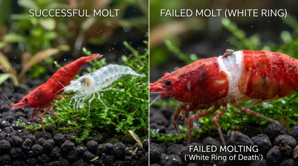
Comparison of successful ecdysis leaving a clear exuviae (left) versus incomplete ecdysis showing the white ring of death cherry shrimp split (right).The physical mechanism that turns a routine shed into a fatal white ring of death cherry shrimp event lies in the fracture geometry of the old exoskeleton during Stage E. During a normal molt, internal hydrostatic pressure splits the old cuticle solely at the dorsal junction between the posterior edge of the carapace and the first abdominal segment, leaving the ventral (underside) cuticle intact.
This intact ventral bridge serves as an essential mechanical fulcrum. When the shrimp executes its forceful “U-bend”—arching its head and tail inward like a coiled spring—the connected underside holds the old shell stationary, providing the leverage required for the shrimp to pop its anterior cephalothorax and long appendages upward and backward out of the dorsal opening.
When water parameter anomalies compromise cuticular elasticity or trigger premature osmotic swelling, the old shell fails to hinge solely at the top of the neck and instead cracks circumferentially (360 degrees) around the entire body. The conspicuous “white ring” is not a pathogen or external growth; rather, it is the pale, unpigmented underlying muscle and newly secreted soft cuticle exposed between the completely severed anterior carapace and posterior abdominal segments.
Because the two halves of the old exoskeleton are now completely disconnected, the mechanical fulcrum vanishes. Each time the shrimp flexes into a U-bend, the separated front and rear shell sections move independently with the body—much like trying to pull off a tight glove that has been torn in half at the wrist. Encased within the rigid anterior carapace helmet, the shrimp cannot free its mouthparts, gills, or walking legs, eventually succumbing to muscular exhaustion, branchial hypoxia, or secondary bacterial infection within 24 to 72 hours.
Other clinical manifestations of incomplete ecdysis include shrimp locked in a permanently curled C-shape post-molt, gravid females prematurely shedding and dropping fertilized egg clutches, or surviving specimens missing walking legs that were amputated inside stuck exuviae.
Pre-Molt Ring vs. White Ring of Death: The 24-Hour Predictive Window
One of the most damaging myths in shrimp husbandry is the belief that any white ring visible on a living shrimp guarantees imminent death, prompting panicked hobbyists to dump chemical additives into the tank or attempt manual shell removal. Strictly defined, the “White Ring of Death” is a post-mortem diagnosis applied after a shrimp has died mid-molt. Every healthy shrimp must develop a separation line at the thoraco-abdominal junction right before ecdysis as internal fluid uptake stretches the arthrodial membrane between the carapace and abdomen.
To test whether visual shell characteristics can predict molt failure before death occurs, empirical 20-case tracking evaluated living shrimp exhibiting visible white rings through completion of their molt cycle. The evaluation tracked two primary morphological anomalies: a large, sustained break (≥ 2 mm) that remained continuously open regardless of posture (whereas normal pre-molt breaks open transiently during grazing and close when the body straightens), and scrunched, crumpled, or irregular edges along the border of the split, which indicate that the shrimp has already attempted and failed to pop out of its shell.
The empirical results overturned conventional hobbyist assumptions: while predictions of successful molts on thin, smooth rings were 90% accurate (9 out of 10 cases), predictions of fatal molt failure on shrimp showing severe warning signs (≥ 2 mm breaks with scrunched edges) were only 40% accurate (4 out of 10 cases). Put differently, 60% of living shrimp displaying severe “problem” rings still managed to molt successfully.
Although a wide, scrunched break elevates the relative risk of molt failure by roughly 3 to 4 times compared to a normal hairline split, it does not guarantee mortality. Crucially for tank management, nearly all observed cases resolved—either via successful shedding or death—within an approximate 24-hour resolution window. Consequently, observing a single shrimp with a questionable ring warrants a 24-hour observation period rather than immediate chemical intervention, whereas multiple shrimp exhibiting wide, scrunched rings confirm systematic cherry shrimp molting failure causes and fix issues across the colony.
| Visual Ring Morphology on Living Shrimp | Edge Texture at Carapace Split | Posture Dependency | Empirical Survival Probability | Diagnostic Classification & Recommended Action |
|---|---|---|---|---|
| Hairline / Thin Split (< 1 mm) | Smooth, clean, uniform margins. | Appears during flexing; disappears when straight. | ~90% Success | Normal Pre-Molt Ring: Healthy Stage D4 apolysis. Do not alter tank parameters. |
| Medium Split (1–2 mm) | Smooth, intact cuticular border. | Partially visible at rest. | ~75–85% Success | Active Ecdysis Preparation: Monitor for 24 hours; verify GH and TDS without panic dosing. |
| Large Sustained Break (≥ 2 mm) | Smooth margins, wide gap. | Continuously visible in all postures. | ~65–70% Success | Elevated Mechanical Risk: Indicates mild osmotic or mineral strain; test GH, KH, and Ca:Mg ratio. |
| Large Break (≥ 2 mm) + Deformed Border | Scrunched, crumpled, or frayed edges. | Permanently wide; appendages sluggish. | ~60% Success / 40% Failure | Stalled Ecdysis: 3–4x failure risk; if seen across multiple shrimp, initiate gradual mineral correction. |
| Complete 360° Split on Immobile/Dead Specimen | Severed anterior/posterior halves; necrotic flesh. | Rigid, curled, or lying on side. | 0% (Terminal / Post-Mortem) | Confirmed White Ring of Death: Execute immediate colony water parameter audit. |
Endocrinology and Biomineralization of Neocaridina Ecdysis
Solving cherry shrimp molting failure causes and fix challenges requires an appreciation of the tightly synchronized neuroendocrine and biomineralization pathways that govern decapod shell renewal. Ecdysis is not a passive mechanical shedding of dead tissue; it is an energy-intensive metabolic cascade regulated by antagonistic hormones and specialized epithelial ion pumps.
The X-Organ/Y-Organ Axis: Molt-Inhibiting Hormone (MIH) and Ecdysteroid Cascades
The initiation and pacing of the crustacean molt cycle are controlled by the interplay between two endocrine structures: the X-organ-sinus gland complex situated within the eyestalks and the paired Y-organs located in the maxillary segment near the base of the antennae. Throughout the intermolt phase (Stage C4), the X-organ secretes Molt-Inhibiting Hormone (MIH).
This peptide neurohormone circulates through the hemolymph and binds to Y-organ receptors, actively suppressing steroid hormone biosynthesis via cyclic nucleotide and intracellular calcium (Ca2+) signaling pathways. When internal growth cues signal that the shrimp has reached the volumetric capacity of its current exoskeleton, X-organ MIH secretion declines.
Freed from MIH antagonism, the Y-organs rapidly synthesize and release ecdysteroids—primarily ecdysone, which peripheral tissues convert into the biologically active steroid hormone 20-hydroxyecdysone (β-ecdysone). Rising ecdysteroid titers in the hemolymph initiate premolt apolysis, stimulate the hypodermis to synthesize the new underlying cuticle, activate enzymatic resorption of calcium from the old shell, and regulate gastrolith formation.
However, if an external environmental stressor—such as an abrupt drop in water temperature or osmotic TDS shock—occurs out of phase with this hormonal timeline, the physical swelling of the body outpaces the ecdysteroid-driven enzymatic weakening of the old cuticle. This hormonal mismatch traps the crustacean inside an unyielding carapace, triggering a fatal white ring of death cherry shrimp molt failure.
Gastrolith Calcium Storage and Amorphous Calcium Carbonate (ACC) Stabilization
Whereas marine crustaceans inhabit seawater containing roughly 400 to 450 ppm of dissolved calcium and obtain approximately 95% of their postmolt calcium directly from the water column, freshwater Neocaridina davidi evolved in habitats with far lower mineral concentrations. Because more than 80% of a crustacean’s total body calcium is deposited within its exoskeleton, discarding the old shell without prior mineral recovery would create an insurmountable postmolt deficit.
To bridge this gap, freshwater shrimp combine high-affinity branchial calcium uptake mechanisms with endogenous mineral recycling. During the 7 to 10 days of premolt, β-ecdysone directs the reabsorption of 20% to 50% of the old cuticle’s calcium carbonate. The shrimp routes a portion into paired, disc-like calcareous concretions in the stomach wall called gastroliths (and phosphate granules in the hepatopancreas), while excess soluble calcium is lost across the gills.
In gravid female cherry shrimp, mature gastroliths can occasionally be observed through the translucent cephalothorax as bright pearly white spots behind the eyes just prior to ecdysis. Storing salvaged calcium as crystalline calcite inside the stomach would render it too slow to dissolve during the urgent 24-to-72-hour postmolt hardening window.
Instead, crustaceans precipitate gastrolith mineral reserves as Amorphous Calcium Carbonate (ACC) and Amorphous Calcium Phosphate (ACP) nanospheres embedded within a chitin-protein nanofiber meshwork. Solid-state nuclear magnetic resonance (NMR) and biochemical analyses reveal that crustaceans prevent ACC from prematurely crystallizing into calcite by binding the nanospheres with specialized matrix phosphoproteins—such as GAP65, GAP10, and GAMP.
This stabilization is further reinforced by phosphorylated intermediates of the glycolytic pathway, specifically phosphoenolpyruvate (PEP) and 3-phosphoglycerate (3PG), as well as citrate-Ca2+-phosphate complexes and magnesium ions. Immediately following ecdysis, the gastroliths collapse into the foregut, where digestive acids dissolve the highly soluble ACC within hours.
According to peer-reviewed crustacean biomineralization research published in PubMed Central, freshwater decapods stabilize resorbed premolt calcium within stomach gastroliths as amorphous calcium carbonate (ACC) bound by specialized matrix phosphoproteins and glycolytic intermediates, enabling rapid cuticular remineralization within hours of ecdysis. This rapid reabsorption quickly calcifies the mandibles and walking legs so the shrimp can resume foraging while extracting the remaining 50% to 80% of its required shell calcium from the aquarium water and diet.
Alkaline Phosphatase, Magnesium Calcite, and Transepithelial Transport
Completing postmolt calcification requires moving massive quantities of divalent calcium (Ca2+) and magnesium (Mg2+) ions from the external freshwater column across the gill, gut, and hypodermal epithelia against an electrochemical gradient. To accomplish this massive transcellular flux without raising free cytoplasmic calcium to cytotoxic levels, epithelial cells utilize specialized active transport systems.
These include Plasma Membrane Calcium-Transporting ATPases (PMCA), Sarcoplasmic/Endoplasmic Reticulum Calcium-ATPases (SERCA), and basolateral Sodium-Calcium Exchangers (Na+/Ca2+). At the cuticular mineralization front—where silicon (as amorphous oligomerized silicic acid) acts as an early nucleation intermediary between polysaccharide-protein complexes and inorganic ions—calcium carbonate precipitates around twisted plywood-like honeycomb matrices of chitin-protein fibers across the epicuticle, exocuticle, and endocuticle.
Within this mineral phase, magnesium (Mg2+) fulfills two non-negotiable biochemical roles. First, it integrates directly into the calcium carbonate lattice to form high-strength magnesium calcite (Mg-calcite) alongside ACC and ACP. Second, magnesium ions act as an essential enzymatic cofactor that modulates alkaline phosphatase (ALP) activity.
When ambient water lacks dissolved magnesium, alkaline phosphatase activity falters, halting calcium deposition into the new exoskeleton even when dissolved calcium is abundant. Simultaneously, external bicarbonate (HCO3–) anions must be present in the water column to support net branchial Ca2+ uptake and precipitate CaCO3 at the cuticular matrix, preventing postmolt collapse and fatal cherry shrimp molting failure causes and fix complications.
Primary Cherry Shrimp Molting Failure Causes and Fix Protocols
When troubleshooting cherry shrimp molting failure causes and fix protocols in home aquariums or breeding setups, aquarists must distinguish between isolated senescence and systemic water-column failures. An aged or previously injured specimen failing a molt in a thriving colony is an isolated event, whereas multiple molting casualties clustered 12 to 48 hours after a water change point directly to osmotic or stoichiometric instability.
Establishing a stable biological baseline requires verifying all core water chemistry metrics against species-specific physiological thresholds. Identifying how individual cations, anions, and osmotic shifts interact enables hobbyists to intervene before cuticular fractures destroy the breeding stock. To establish a rock-solid baseline before adjusting individual ions, aquarists should verify all core metrics against the comprehensive cherry shrimp water parameters guide.
Osmotic Shock, TDS Creep, and the Premature Ecdysis Trigger
The primary trigger of acute white ring of death cherry shrimp outbreaks is osmotic shock resulting from large, rapid water changes with mismatched Total Dissolved Solids (TDS), General Hardness (GH), or temperature. Because freshwater dwarf shrimp maintain a pressurized internal hemocoel encased in chitin, sudden shifts in ambient solute concentrations immediately distort internal fluid dynamics.
When an aquarist pours softer, lower-TDS water into a tank during a heavy water change, water rushes inward across the semi-permeable gill membranes via osmosis. This osmotic surge inflates the shrimp’s body prematurely, bursting the old exoskeleton open days before the Y-organ ecdysteroid cascade has resorbed internal minerals or synthesized the new soft cuticle.
Because the old cuticle has not yet been enzymatically weakened along the dorsal hinge, the hydrostatic expansion rips the shell circumferentially around the entire waist. This mechanical tear guarantees a fatal incomplete molt within 12 to 48 hours.
This mortality spike is frequently exacerbated by evaporative TDS creep. As pure water evaporates from the aquarium surface, dissolved calcium, magnesium, sulfates, and trace salts remain behind, steadily concentrating total mineral density.
If an aquarist tops off evaporated water with mineralized tap water instead of zero-TDS RO/DI water, General Hardness climbs relentlessly. A subsequent routine water change then subjects the shrimp to a violent downward osmotic drop. Automating evaporation replenishment via a calibrated aquarium auto top off setup filled strictly with 0-TDS RO/DI water prevents evaporative solute drift, while implementing the membrane-flushing protocols in the osmotic shock prevention guide prevents high-TDS permeate bursts from entering the reservoirs. Furthermore, when acclimating newly acquired shrimp or refilling tanks after maintenance, applying the physiological principles of drip acclimation vs plop and drop is essential; invertebrates must never undergo raw plop-and-drop transfers across unbridged osmotic gradients.
Hypo-Mineralization (Low GH) vs. Hyper-Calcification (Excess GH and Low pH)
General Hardness (GH)—which quantifies dissolved divalent cations, predominantly Ca2+ and Mg2+, where 1 dGH equals 17.86 ppm as CaCO3—governs the physical thickness and structural rigidity of the crustacean exoskeleton. Deviations outside the optimal 6 to 10 dGH (107 to 179 ppm) window for Neocaridina davidi produce two distinct cuticular failure modes.
In hypo-mineralized water (GH < 6 dGH), which commonly occurs when hobbyists use un-remineralized RO water or naturally soft municipal tap water, shrimp cannot extract sufficient mineral ions during the critical 24-to-72-hour postmolt hardening window. Consequently, the newly synthesized cuticle remains abnormally thin, soft, and flexible.
When the subsequent molt cycle arrives, the malleable shell stretches and tears raggedly instead of snapping cleanly at the dorsal seam. This leaves the animal fatally trapped inside a rubbery, collapsed cuticular tube.
Conversely, hyper-calcification (GH > 14 to 15 dGH) develops when aquarists overdose calcium supplements, accumulate mineral creep, or maintain colonies in exceptionally hard liquid-rock water. Excessive calcium deposition renders the old exoskeleton overly thick, dense, and inflexible.
Compounding this mechanical barrier, chronically acidic water (pH < 6.5) in Neocaridina aquariums alters the calcium weight distribution within the cuticle, disrupting the cuticular Ca:Mg balance and increasing shell density. When the shrimp attempts to flex out of a hyper-calcified shell, the muscular U-bend cannot generate sufficient leverage to rupture the rigid dorsal joint. The brittle armor snaps 360 degrees around the middle or exhausts the animal to death. If municipal tap water exceeds 14 dGH, aquarists should consult the dilution calculations on how to lower aquarium GH in freshwater tanks to blend pure RO water safely into their weekly maintenance routine.
The Calcium-to-Magnesium (Ca2+:Mg2+) Stoichiometric Imbalance
Clinical aquaculture data from NCBI studies on crustacean calcium and magnesium metabolism confirm that over 80% of total body calcium resides in the exoskeleton and that dissolved magnesium ions directly regulate postmolt shell formation by modulating alkaline phosphatase activity and integrating into the cuticle as structural magnesium calcite.
Even when a liquid drop test confirms an apparently optimal 8 dGH reading, colonies frequently suffer recurring white ring of death cherry shrimp mortalities if the underlying Calcium-to-Magnesium (Ca2+:Mg2+) mass ratio is skewed. Standard GH titration kits measure total divalent cations collectively, making it impossible to detect whether an 8 dGH reading represents balanced minerals or pure calcium with near-zero magnesium.
Magnesium ions are physiologically indispensable for cuticular synthesis. They co-precipitate to form high-strength magnesium-calcite in the endocuticle and serve as an obligate enzymatic cofactor that activates alkaline phosphatase (ALP) for transcellular calcium absorption.
When aquarists rely solely on pure calcium supplements—such as calcium carbonate powders, eggshells, or cuttlebone—in magnesium-deficient water, the Ca:Mg ratio easily exceeds 6:1 or 10:1. This induces functional magnesium starvation, stalling cuticular mineralization despite high total calcium levels.
For Neocaridina davidi, the optimal mass concentration ratio is 3:1 to 2:1 Calcium-to-Magnesium (representing a 4:1 ratio of total GH to magnesium). Preserving this balance requires a dedicated shrimp remineralizer containing balanced salts or targeted Epsom salt (MgSO4·7H2O) supplementation.
Carbonate Depletion (Low KH), Active Soils, and Branchial ATP Exhaustion
Carbonate Hardness (KH) quantifies dissolved carbonate (CO32-) and bicarbonate (HCO3–) anions, which buffer pH and provide inorganic carbon for cuticular CaCO3 precipitation. In biological systems, autotrophic nitrification continuously consumes alkalinity, exhausting 7.14 mg of carbonate buffer (as CaCO3) for every 1.0 mg of ammonia-nitrogen oxidized:
2H+ + 2HCO3– → 2H2O + 2CO2
Alkalinity loss accelerates rapidly when hobbyists house hard-water Neocaridina over active humic buffering aquasoils. Active substrates possess a high Cation Exchange Capacity (CEC) that strips Ca2+ and Mg2+ while releasing H+ ions that neutralize bicarbonate toward 0 dKH.
When rejuvenating depleted planted substrates in a Neocaridina tank, aquarists should follow the protocols on how to recharge old aquarium soil using deeply buried root tabs (placed 1.5 to 2 inches deep) rather than capping the tank with fresh, unbuffered active soil that triggers immediate GH and KH collapse.
When KH drops below 2 dKH (35.7 ppm), pH stability collapses and branchial calcium uptake stalls. Under low-KH, acidic conditions, crustaceans must upregulate branchial Carbonic Anhydrase (CA) enzymes, expending massive amounts of cellular ATP simply to regulate internal hemolymph pH.
This severe energetic drain deprives the shrimp of the metabolic fuel required for the physical ecdysis jump. To maintain carbonate levels between 2 and 5 dKH, aquarists should avoid household baking soda (NaHCO3)—which drives cumulative sodium toxicity—and instead utilize potassium bicarbonate (KHCO3) or crushed coral media.
To restore carbonate reserves between 2 and 5 dKH without harming the colony, aquarists should implement the chemical strategies in our guide on how to safely raise low kh in freshwater aquarium systems.
Nutritional Deficiencies: Cholesterol Ecdysteroid Precursors, Protein Myths, and the Iodine Fallacy
While water-column minerals provide roughly 75% of external shell calcification, diet dictates whether shrimp can synthesize molting hormones and build the organic chitin-protein framework. Because crustaceans cannot synthesize sterol rings de novo, they depend on dietary cholesterol, phytosterols, and phospholipids for Y-organ ecdysteroid synthesis. Diets consisting solely of low-grade flake foods or plain vegetables induce sterol deficiencies that interrupt hormonal ecdysone cascades.
Furthermore, hobbyists must discard the common myth that dietary protein causes shell overgrowth and triggers the white ring of death cherry shrimp. Controlled nutritional studies confirm that dwarf shrimp thrive on diets containing 40% to 47% crude protein without molting defects.
Because the crustacean exoskeleton consists of 30% to 40% structural protein, 20% to 30% chitin, and 30% to 50% calcium carbonate, eliminating protein deprives the new cuticle of essential amino acids and triggers colony cannibalism. Providing blanched kale delivers an optimal 1:1 balance of bioavailable calcium (33 mg/100g) and magnesium (33 mg/100g), while leaving discarded exuviae in the tank allows shrimp to naturally recycle 20% to 25% of their mineral budget.
Finally, evidence-based husbandry requires rejecting the freshwater iodine supplementation fallacy. While marine crustaceans utilize dissolved iodine, freshwater Neocaridina obtain adequate trace iodine from commercial feeds, spirulina, and botanicals.
Because standard test kits cannot measure iodine and the margin between therapeutic trace exposure and invertebrate toxicity is exceptionally narrow, liquid iodine dosing routinely induces fatal poisoning. Similarly, cultivators must maintain zero tolerance for copper (Cu2+), which has a 96-hour LC50 of just 0.0313 mg/L, and quarantine non-tissue-culture plants to eliminate residual pesticides that sabotage ecdysis.
| Parameter / Factor | Neocaridina davidi (Cherry Shrimp) Target | Caridina cantonensis (Bee Shrimp) Target | Pathological Molt Failure Threshold | Primary Physiological Impact on Ecdysis |
|---|---|---|---|---|
| General Hardness (GH) | 6–10 dGH (107–179 ppm) | 4–6 dGH (71–107 ppm) | < 6 dGH (Soft shell) or > 14 dGH (Rigid armor) | Governs Ca2+ and Mg2+ availability for postmolt Mg-calcite endocuticle mineralization. |
| Carbonate Hardness (KH) | 2–5 dKH (36–89 ppm) | 0–2 dKH (0–36 ppm) | < 2 dKH (pH crash / CA drain) or > 8 dKH | Buffers ambient pH and supplies HCO3– anions required for cuticular CaCO3 deposition. |
| Calcium : Magnesium Ratio | 3:1 to 2:1 (by mg/L mass) | 3:1 to 2:1 (by mg/L mass) | > 6:1 (Mg2+ starvation) or < 1:1 | Mg2+ activates alkaline phosphatase to enable transcellular Ca2+ uptake into new shell. |
| Total Dissolved Solids (TDS) | 150–250 ppm (Max 300 ppm) | 100–180 ppm | Sudden swing > 20–30 ppm during water change | Regulates internal osmotic pressure; rapid downward drops trigger premature shell splitting. |
| pH Level | 6.8–7.6 (Tolerable 6.5–8.0) | 5.5–6.6 (Active soil buffered) | < 6.5 (Alters shell Ca density) or rapid ΔpH > 0.5 | Acidic drift in Neocaridina setups alters cuticular Ca:Mg ratios and exhausts cellular ATP. |
| Water Temperature | 20–25°C (68–77°F) | 18–23°C (65–74°F) | > 28°C (Hypoxic rapid molts) or ΔT > 2°C drop | Controls metabolic molt pacing; cold water change additions trigger immediate stress ecdysis. |
| Ammonia / Nitrite / Nitrate | 0 ppm / 0 ppm / < 10 ppm | 0 ppm / 0 ppm / < 5 ppm | NH3 > 0, NO2– > 0, NO3– > 20 ppm | Chronic nitrate exposure halts juvenile development and triggers adult molting mortality. |
| Copper (Cu2+) & Pesticides | 0.000 mg/L (Strict Zero) | 0.000 mg/L (Strict Zero) | > 0.0313 mg/L (96-hr LC50 lethal toxicity) | Potent branchial toxin and neurotoxin; directly inhibits ecdysis and triggers mass die-offs. |
Mathematical Remineralization and Emergency Colony Rescue Protocols
When multiple shrimp exhibit stalled molts or confirmed white ring of death cherry shrimp fatalities, aquarists must resist the urge to perform reactionary parameter surgery. Dumping unmeasured mineral powders directly into the display tank or executing massive emergency water changes only amplifies osmotic trauma and accelerates mortality.
Instead, precision titration testing and displacement-adjusted gravimetric dosing should be calculated using the interactive aquarium calculator and parametric log to account for exact net working water volume after hardscape and substrate displacement. Systematically isolating mineral deficits provides a reliable foundation for executing targeted cherry shrimp molting failure causes and fix strategies without destabilizing water chemistry.
Calculating Calcium and Magnesium Deficits Using Titration Telemetry
To determine whether an apparently healthy GH reading conceals a fatal Ca2+:Mg2+ imbalance, aquarists can combine a standard freshwater GH titration drop test with a modified API Calcium liquid test. By filling the test vial with 10 mL of freshwater aquarium water, adding 20 drops of Calcium Reagent Bottle 1, and counting the drops of Reagent Bottle 2 required to shift the solution from pink to pure blue, each drop corresponds to 10 mg/L Ca2+ (or 5 mg/L resolution if water and reagent volumes are doubled to 20 mL and 40 drops).
Because total General Hardness in ppm CaCO3 (GHppm = dGH × 17.86) equals the sum of dissolved calcium and magnesium converted to calcium carbonate equivalents (where 1 mg/L Ca2+ = 2.497 ppm CaCO3 and 1 mg/L Mg2+ = 4.118 ppm CaCO3), the exact dissolved magnesium concentration ([Mg2+] in mg/L) is isolated using the stoichiometric balance equation:
[Mg2+] = ((dGH × 17.86) − (2.497 × [Ca2+])) / 4.118
For example, if a 38-Liter (10-gallon) Neocaridina aquarium measures 7 dGH (125.02 ppm CaCO3) and 40 mg/L Ca2+, substituting these values reveals an actual magnesium concentration of only 6.10 mg/L Mg2+:
[Mg2+] = ((7 × 17.86) − (2.497 × 40)) / 4.118 = (125.02 − 99.88) / 4.118 = 6.10 mg/L
Dividing 40 mg/L Ca2+ by 6.10 mg/L Mg2+ exposes a severely skewed Ca:Mg ratio of 6.55:1, explaining why the colony experiences molting failures despite an apparently normal 7 dGH reading. To correct this deficit toward an optimal 2:1 or 3:1 ratio without overshooting total GH, the exact mass in grams of Epsom salt (MgSO4·7H2O, containing 9.86% elemental Mg) or gypsum (CaSO4·2H2O, containing 23.28% elemental Ca) is calculated via the volumetric dosing equation:
Massadditive (g) = ((Ctarget − Ccurrent) × Vnet (L)) / (Purity Fraction × 1000)
Step-by-Step Emergency Colony Stabilization Protocol and Checklist
When an aquarist detects stalled molts or confirmed post-mortem white ring of death cherry shrimp casualties, colony rescue depends on stabilizing osmotic gradients while slowly correcting mineral stoichiometry over multiple days. First and foremost, manual extraction with tweezers must never be attempted.
Because the newly formed cuticle beneath the split carapace is paper-thin and uncalcified, attempting to pry or lift the stuck shell crushes the cephalothorax, rips branchial lamellae, or amputates limbs, resulting in a 99% mortality rate. Leaving a living shrimp undisturbed in a dimly lit, moss-rich tank allows the natural 24-hour resolution window to play out, where 60% of even severely scrunched pre-molt breaks resolve successfully on their own.
Simultaneously, the aquarist should test both tank water and replacement tap/RO water for GH, KH, pH, TDS, ammonia, nitrite, and nitrate using liquid titration kits. If GH is below 6 dGH or the Ca:Mg ratio is skewed, mineral corrections must be pre-dissolved into replacement water and dripped in slowly via an airline tube.
To prevent secondary osmotic shock, GH must never be raised by more than 1 to 2 dGH per 24-hour period, and KH must never be raised by more than 1 dKH (17.86 ppm) per 24-hour period. Placing passive calcareous buffers—such as boiled cuttlebone, crushed coral in a filter media bag, Wonder Shells, or montmorillonite clay—provides a gentle, self-regulating mineral release while the water column stabilizes.
Rapid-Reference Triage and Maintenance Checklist
- Zero Manual Interference: Leave living shrimp with visible white rings completely untouched for 24 hours; never use tweezers to pull stuck shells.
- Liquid Titration Audit: Measure GH (6–10 dGH), KH (2–5 dKH), TDS (150–250 ppm), pH (6.8–7.6), and Calcium/Magnesium (3:1 to 2:1 ratio) before adding any chemical supplements.
- Strict Daily Adjustment Ceiling: Limit hardness corrections to a maximum of ≤ 1–2 dGH/day and ≤ 1 dKH/day using pre-dissolved shrimp remineralizer salts (GH/KH+).
- 10% Drip-Refill Water Changes: Replace large bucket pours with 10–15% weekly water changes matched within ±1 dGH, ±10–20 ppm TDS, and ±0.5°C, dripped into the tank at 1 to 3 drops per second.
- 0-TDS Evaporation Top-Offs: Replenish evaporated water exclusively with 0-TDS RO/DI water to eliminate chronic TDS and hardness creep.
- Sterol, Protein & Mineral Nutrition: Rotate cholesterol-rich shrimp pellets (35–40% protein) with blanched organic kale, spinach, and mulberry leaves, and leave all shed exoskeletons (exuviae) inside the tank for mineral recycling.
Conclusion: Eliminating the White Ring of Death Cherry Shrimp
Eliminating the white ring of death cherry shrimp pathology requires moving beyond reactionary chemical dosing to establish predictable osmotic and stoichiometric stability. While a complete 360-degree fracture across the thoraco-abdominal junction destroys the ventral fulcrum needed for the hydrostatic U-bend escape, empirical case tracking proves that thin pre-molt separation lines are a normal feature of Stage D4 apolysis and that 60% of living shrimp displaying even wide, scrunched breaks successfully shed their armor within 24 hours.
By maintaining General Hardness at 6 to 10 dGH with a verified 3:1 to 2:1 Calcium-to-Magnesium ratio to support alkaline phosphatase activation, preserving 2 to 5 dKH to prevent branchial Carbonic Anhydrase ATP exhaustion, topping off evaporation strictly with 0-TDS RO/DI water, and executing 10% airline-dripped water changes alongside a sterol- and protein-balanced diet, aquarists create a homeostatic environment where Neocaridina davidi colonies molt and reproduce effortlessly across generations.
Frequently Asked Questions (FAQs)
Does a white ring on a living cherry shrimp always mean it will die?
No. A thin, smooth white separation band between the posterior margin of the carapace and the first abdominal segment is a normal anatomical phase of premolt apolysis as the shrimp absorbs water to expand its body prior to ecdysis. Even when a living shrimp exhibits a wide break (≥ 2 mm) with scrunched cuticular edges, empirical case tracking demonstrates that approximately 60% still complete their molt successfully within 24 hours. The term white ring of death cherry shrimp is strictly a post-mortem diagnosis applied after the molt has failed and the shrimp has perished.
Can aquascaping tweezers be used to help a cherry shrimp stuck in its old shell?
Manual extraction with tweezers should never be attempted. The newly secreted cuticle underneath the old exoskeleton remains paper-thin and uncalcified for 24 to 72 hours following apolysis. Attempting to pull or pry the stuck carapace manually tears delicate branchial tissue, amputates walking legs, or crushes internal organs, resulting in a 99% fatality rate. Leaving the specimen undisturbed in calm, well-oxygenated water offers its highest probability of natural survival.
Why are cherry shrimp dying with the white ring even when GH is 8 dGH?
When molting failures persist despite an optimal 6 to 10 dGH reading, investigating cherry shrimp molting failure causes and fix strategies typically uncovers one of three hidden factors: (1) a skewed Calcium-to-Magnesium (Ca2+:Mg2+) ratio where magnesium is too depleted to activate alkaline phosphatase for shell mineralization; (2) osmotic shock triggered by pouring in large water changes that differ in TDS or temperature; or (3) dietary cholesterol/sterol deficiency, which deprives the Y-organ of the lipid precursors required to synthesize ecdysteroid molting hormones.
Should shed shrimp exoskeletons (molts) be removed from the aquarium?
No. Discarded exoskeletons (exuviae) are sterile, non-polluting mineral structures composed of calcium carbonate, magnesium calcite, chitin, and structural proteins. Healthy shrimp graze on and consume the empty shells within 24 to 48 hours to reclaim 20% to 25% of their required calcium and trace elements. Shed shells should only be removed if the aquarium is undergoing active treatment for an external scutariellid parasite infestation.
Does dosing liquid iodine cure cherry shrimp molting failures?
No. Scientific evaluation confirms that deliberate liquid iodine supplementation in freshwater dwarf shrimp aquariums is unnecessary and potentially lethal. Unlike marine systems, freshwater Neocaridina acquire ample trace iodine from quality shrimp pellets, spirulina, decomposing botanicals, and commercial remineralizer salts. Because standard freshwater aquarium test kits cannot measure iodine concentrations and excess iodine is acutely toxic to crustaceans, unmeasured dosing frequently triggers colony-wide mortality.
